
 Search
 Global
ProSound F75 Premier/CV
FIT to your specialty
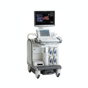
The ProSound F75 Premier/CV is a high-end, diagnostic ultrasound system which provides excellent performance in a variety of clinical applications and is comfortable and easy to use.
The ProSound F75 Premier/CV system has been designed with our "FIT" ethos (Facilitate workflow; Investment return; True diagnostic) at its heart and can be tailored to the needs of the clinician. The ProSound F75 Premier/CV reduces burden on the examiners and patients, assures and maintains versatility at a high dimension, reduces patient dependency by increasing the level of diagnostic ability, and improves productivity of routine examinations by accuracy and efficiency.
F: Facilitate workflow Making routine examinations more comfortable
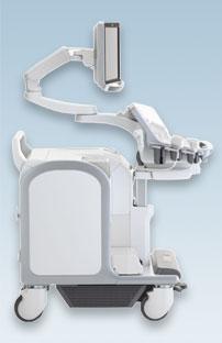
The ProSound F75 Premier/CV acknowledges the needs of performing examinations in a natural posture without stress or discomfort. The monitor and operation panel have a wide range of movement, which enables reduction of the operator's muscle load up to 20%. Another factor is automation of operation, to make examinations flow smoother. The ProSound F75 Premier/CV allows simple operation so the operator can concentrate on the examination in comfort.
- NaturalErgonomics™
- Widely height adjustable console panel
- Freely placeable viewing monitor
- High Definition (HD) Wide IPS-pro viewing monitor
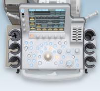
- Large IPS-pro touch panel with intuitive 8 rotary encoder
- Routine exam assistance
- Quick Setter™
- Image Optimizer™
I: Investment return for the hospital management and society
The ProSound F75 Premier/CV covers numerous applications in one console, and offers wide variety of probes, measurement/report package, and many other factors. Affordable cost of ownership is made possible due to the reliable system design and easy-to-upgrade software. With simple and intuitive operation, new users can easily learn to operate the system.
- High degree of versatility
- Variety of transducers include TEE, TV, TR
- Networking, measurement and report package
- Compact console fits any clinical setting
- Reduce TCO (total cost of ownership)
- Acquisition - short learning time for sonographer to operate the system, compatibility of transducers
- Operation - reliability/low running cost
- Long term - Software backend, low future upgrade cost
- EcoEcho - Environmental performance
- RoHS - EU directive
- WEEE - EU directive
- Material efficiency / recycle care
T: True diagnostics reduce the patient dependency
By combining all of our cutting-edge technologies, the ProSound F75 Premier/CV performs high spatial resolution. The combination of the CPWG+ (compound pulse wave generator, the new front-end processor) and Full Aperture Apodization (uses all transducer elements of a probe) perfects focus throughout the entire image. This technology enhances the efficiency of examinations, and makes benefit both examiners and patients.
- CPWG+ Frontend
- FAA: Full Aperture Apodization
- TAT: Tissue Adaptive Technology
- CTT: Clear Transmission Technology
- SmartProbe™
- Broadband Harmonics (BbH)
- Spatial Compound Imaging+ (SCI+)
- AIP: Adaptive Imaging Processing
- Edge Optimizer
- eFLOW+
- Trapezoidal Scan
- EFV: Extended Field of View
- Real-time 3D (4D)
- Contrast Echo (CE)
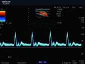
Carotid artery with Compound Gated Pulse Wave Doppler |
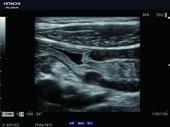
Digestive tract with outstanding image quality |
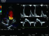
Dual Doppler enables observation of 2 Doppler waveforms at the same heartbeat |
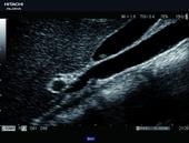
Gallbladder polyp in high quality zoom |
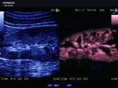
Kidney with eFLOW+x Flow Emphasis x DDD |
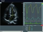
Quantification of intracardiac volume - 2DTT |
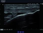
Tibial tuberosity with AIP HI REZ and SCI+ |
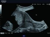
Uterine cervix in a wide field of view |
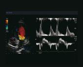
Dual Gate Doppler: Combination of PW and TDI allows simulaneous evaluation of wall motion and hemodynamics |
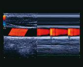
Wave Intensity (Ventriculo-Arterial Coupling): WI is a new hemodynamic index which is potentially useful for analyzing interaction of the cardiovascular system (heart and blood vessels) |
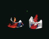
3D Zoom mode (Flow) |
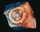
Dynamic Volume Processing |
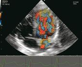
VFM - Vector Flow Mapping - Evaluation of cardiac blood flow motion |
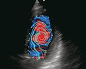
VFM - Apical four-chamber view - blood flow streamlines in diastole |
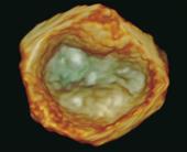
3D zoom Clear structures of the mitral valve |
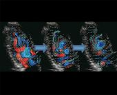
VFM - Flow Patterns in an apical long axis view - Rapid inflow phase, slow inflow phase 1, slow inflow phase 2 (left to right) |