
 Search
 Global
ProSound Alpha 7
Powerful, friendly and compact ultrasound system
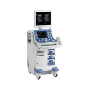
The ProSound Alpha 7 is a diagnostic ultrasound system that contradicts the thought that high-performance systems are large. It inherits the proven technologies and functions of our high-end products, and offers outstanding mobility thanks to being one of the smallest size in its class. The system is easily transportable to ensure high performance throughout the hospital.
The Broadband Harmonics realizes high sensitivity that is comparable to fundamental imaging even with Harmonic Echo imaging. eFLOW features enhanced spatial resolution for greater detail of blood flow information. The 3D Automated Volume Measurement (AVM) requires no manual tracing for accurate 3D volume calculation. Contrast Harmonic Echo (CHE) is compatible with all high-, medium- and low-sound pressure contrast agents.
Significant effort was exerted to create a system that would alleviate user fatigue and increase patient throughput by applying universal design. The use of eco-friendly materials, low power consumption and low noise design makes ProSound Alpha 7 an environment friendly system.
By packing Broadband Harmonics™, high-quality imaging with high-performance HST+ probes, analysis functions, and Real-time 3D/4D into a very small console, the ProSound Alpha 7 enables examinations that rival those of premium systems.
Broadband Harmonics™
Harmonic imaging by Broadband Harmonics™ technology provides sensitivity and resolution comparable to those of fundamental imaging. In addition to reducing side lobes and multiple echoes, more detailed image information and much higher penetration in deeper areas are achieved.
eFLOWCompared with conventional blood flow display methods, eFLOW features enhanced spatial and time resolutions for greater detail. The ProSound Alpha7 detects all flows (in narrow vessels with very low velocity flows) as well as high velocity flows (in adjacent large vessels) and automatically displays them without neither overlapping tissue images/flows nor user’s PRF adjustments. Moreover, by assigning separate colours to flows moving toward and away from the transducer, it is also possible to separately display opposite flows traveling in parallel.
Image Optimizer
The image optimizer enhances the brightness, the horizontal gain and lateral gain of the entire B-mode image. The user is freed from frequent image adjustments during examination, resulting in enhanced examination efficiency.
Adaptive Image Processing (AIP): A revolutionary feature for speckle noise reductionAIP clearly displays differences in tissues, reducing speckle noise while maintaining the frame rate. Simultaneously, it displays outlines more clearly by selectively emphasizing boundaries.
Advanced 3D/4D Functions- Multi-planar Reconstruction (MPR)
- 3D Automated Volume Measurement (AVM)
- Multi Slice Imaging (MSI)
- Flow 3D
User-friendly Universal Design
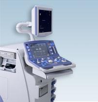
- The control panel swivels and is height adjustable
- High-resolution flat panel LCD monitor with vertical adjustments on flexible arm
- User-customizable panel switches
- Multi-function controls
Environmentally friendly
About 40% less power consumption than a conventional model.
CompactLight and compact system, allowing for excellent portability.
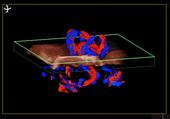
3D display of blood flow in the umbilical cord using Flow 3D |
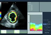
A-SMA - Automated Segmental Motion Analysis |
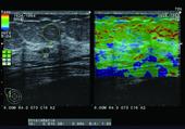
Breast with RTE Real-time Tissue Elastography |
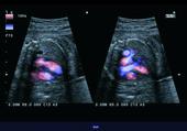
DSD-Dynamic Slow-motion Display |
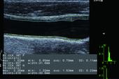
IMT - Automated Intima-media Thickness Measurement |
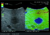
Intraoperative - Metastatic hepatic cancer with RTE Real-time Tissue Elastography |
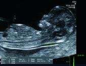
AutoNT - Automated Nuchal Translucency Measurement |
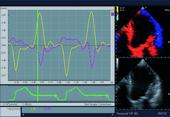
TDI - Tissue Doppler Imaging Analysis |
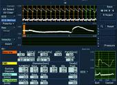
WI - Wave Intensity |